Description
Dermatoscopy and Skin Cancer, second edition, is a handbook to help dermatologists, dermatoscopists and GPs easily differentiate between benign and malignant tumours, leading to fewer unnecessary biopsies and earlier treatment of cancers.
Based around two easy to follow algorithms, ‘Chaos and Clues’ and ‘Prediction without Pigment’, the book shows all dermatoscope users how to confidently diagnose skin lesions earlier and with greater precision.
In addition, this handbook provides coverage of:
- the microanatomy of the skin
- specimen processing and histopathology
- the language of dermatoscopy to help name and define structures and patterns
- approaches to skin examination and photodocumentation
- revised pattern analysis as an additional diagnostic algorithm
- dermatoscopic features of common and significant lesions.
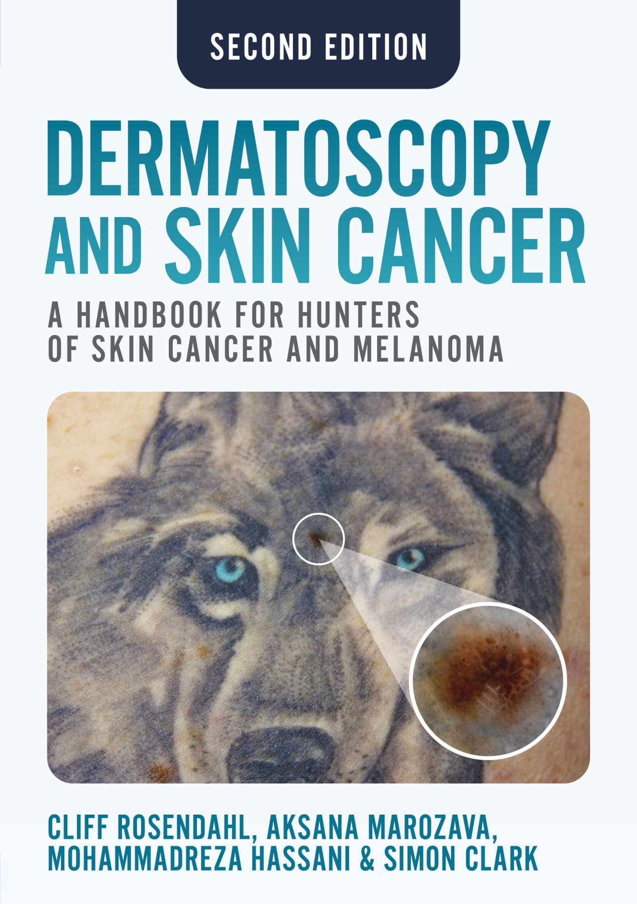
Using hundreds of high quality images, the authors provide a detailed algorithmic approach to assessing the skin; an approach that has been successfully taught to thousands of doctors around the world.
From Doody’s reviews, December 2023
“Many dermoscopy books exist; some are too pedantic and explain concepts with dermatoscopic jargon, while others purport to simplify the learning process but quickly succumb to the same criticism. Most are replete with abnormal looking lesions, but fall short on including examples of normal variations. This book delivers what it promises. I definitely recommend it as the first reference for mastering diagnosis of skin lesions with a dermatoscope.” – 4 stars!
Preface to the second edition; Foreword; Abbreviations
Chapter 1: Introduction to dermatoscopy
1.1 Why use a dermatoscope?
1.2 What is a dermatoscope?
1.3 Colours in dermatoscopy
1.4 Differences between polarised and non-polarised dermatoscopy
1.5 Uses of dermatoscopy for conditions other than tumours
Chapter 2: Skin – the organ
2.1 Skin as an organ
2.2 Embryology of skin
2.3 The microanatomy of skin
Chapter 3: Dermatopathology for dermatoscopists
3.1 Introduction
3.2 Tissue and context: the two pillars of reliable skin cancer diagnosis
3.3 From the scalpel to the microscope: the specimen journey
3.4 The histology of normal skin
3.5 Core dermatopathology terminology
3.6 Neoplastic lesions: dermatoscopic–histological correlation
3.7 Clinicopathological collaboration in skin cancer diagnosis
Chapter 4: The language of dermatoscopy: naming and defining structures and patterns
4.1 The evolution of terminology for dermatoscopic structures and patterns
4.2 Revised pattern analysis of lesions pigmented by melanin
4.3 Patterns in revised pattern analysis
4.4 The process of revised pattern analysis
4.5 Revised pattern analysis applied to lesions with white structures
4.6 Revised pattern analysis applied to lesions with orange, yellow and skin-coloured structures
4.7 Revised pattern analysis – a diagnostic algorithm
4.8 An aide-memoire for revised pattern analysis of pigmented skin lesions
4.9 Revised pattern analysis applied to vessel structure and patterns
4.10 The cognition of dermatoscopy
Chapter 5: The skin examination
5.1 The skin check consultation
5.2 Photo-documentation
5.3 Patient safety: tracking specimens and self-audit
5.4 The lives of lesions
Chapter 6: Chaos, Clues and Exceptions: a decision algorithm for pigmented skin lesions
6.1 'Chaos, Clues and Exceptions'
6.2 Chaos
6.3 Clues
6.4 Exceptions
Chapter 7: Prediction without Pigment: an algorithm for non-pigmented skin lesions
7.1 'Prediction without Pigment'
7.2 'Prediction without Pigment': Part 1
7.3 'Prediction without Pigment': Part 2
7.4 Conclusion
Chapter 8: Melanoma
8.1 What is a melanoma?
8.2 Melanoma subtypes
8.3 Melanomas with adverse outcomes
8.4 Metastatic melanoma
Chapter 9: Melanocytic naevi
9.1 Melanocytic naevi, pigmented and non-pigmented
9.2 Dysplastic naevus
Chapter 10: Basal cell carcinoma, benign and malignant keratinocytic lesions, distinguishing flat pigmented facial lesions, dermatofibroma, vascular and other lesions
10.1 Basal cell carcinoma: pigmented and non-pigmented
10.2 Benign keratinocytic lesions
10.3 Actinic keratosis and squamous cell carcinoma in situ
10.4 Squamous cell carcinoma and keratoacanthoma
10.5 Distinguishing flat pigmented facial lesions
10.6 Dermatofibroma and dermatofibrosarcoma protuberans
10.7 Haemangioma and other vascular lesions
10.8 Merkel cell carcinoma
10.9 Atypical fibroxanthoma
10.10 Adnexal tumours
10.11 Neurofibroma
10.12 Molluscum contagiosum
10.13 Cutaneous lymphoma
10.14 Kaposi sarcoma
Chapter 11: Photographic technology as a diagnostic tool in melanoma management
11.1 Utilisation of photographic technology in skin lesion diagnostics
11.2 Serial digital dermatoscopic imaging
11.2.1 Serial digital dermatoscopic imaging – targeted and random
11.2.2 Selection by serial digital dermatoscopic imaging – a promising alternative to random lesion selection (SDDI – TBPD)
11.2.3 Serial digital dermatoscopic imaging – which lesions should be excised?
11.3 Total body photography
11.3.1 Total body photography used as a baseline during skin examination
11.3.2 Serial total body photography with detection software
Index
Five-star reviews:
Outstanding skin cancer diagnosis book for beginners and experts alike "This book is based on some 20,000 skin lesion cases which Professor Rosendahl personally diagnosed, photographed and treated in his Brisbane clinic, and analysed through the SCARD data base which he helped create. That is serious data from real life, giving this book great authority. I had the privilege of studying in the author's clinic, and as a doctor of 40 years, 20 years working with skin tumours, I know a real expert when I see one at work.
"Cliff is passionate about skin cancer diagnosis, and teaching it to others. Highly regarded as an original researcher, he teaches skin lesion recognition all over the world, including places like Iran, Turkey and Ukraine as well as Australia, New Zealand and Western Europe. He worked with Viennese dermatoscopy superstar Professor Harald Kittler to develop the 'Chaos and Clues' and 'Prediction without Pigment' algorithms which run through the whole book.
"This modern diagnostic approach builds on previous knowledge and is objectively as diagnostically accurate as older diagnostic methods, but is quicker to learn and easier to teach. Moreover, the method uses an objective, geometric , descriptive terminology for lesion patterns and clues, which translates into non-English languages more easily than the older metaphorical terminology (very necessary since skin cancer and dermoscopy are global) and gives a more reproducible way of sharing data for research.
"Strongly recommended for all skin lesion diagnosticians from nurse to professor, beginners and advanced will all get something from this book. Very inexpensive too!" Amazon reviewer
(Declaration of interest: SH received generous hospitality and tuition from Cliff Rosendahl when visiting Brisbane, and contributed a foreword to the book.)
Superb "The most informative and accessible book I have read on this subject. Great illustrations and clear, informative, relevent text on skin structure, histopathological correlations of dermatoscopy and methodology for dermatoscopy along with excellent images. Tremendous value for money." Amazon reviewer
Beautiful examples of clinico-dermoscopic-pathologic correlation "Rosendahl and Marozava’s Dermatoscopy and Skin Cancer is a great resource for both beginner dermoscopists and those wishing to advance their skills. Illustrations combining clinical, dermoscopic and histopathologic images emphasise the correlations between these three critical modalities and are particularly useful for understanding and identifying the whole range of skin cancers and their stimulants." Amazon reviewer
The Best Dermatoscopy informative book going! World class standard reference medical text "This very high quality book co-written by one of the world's leading light experts (with his associate - Aksana) in the field of skin cancer detection by dermatoscopy. Beautifully presented by a very articulate Professor of Medicine author using his own dermatoscopic photo's. The book and its succinct content is also very well organized and indexed.
Every Skin Cancer Medical Doctor must have this book on his / her top shelf as an excellent resource and reference as is my copy, in our quest to master early detection and zero tolerance approach to skin cancer"
Amazon reviewer
A very useful book "This book is useful and "solid". No frills. Much practical experience, excellent teaching organization. There is everything you need to know not only for the family doctor, but also for an experienced dermatoscopist.
Recommended as a fundamental text for the topic." Amazon reviewer
[The book] is laid out with clear, basic language that
students of the skin at any level will benefit from. This handbook starts with
a review of basic dermatoscopic techniques and concepts, followed by a basic
science review of the anatomy, embryology, histology, and pathophysiology of
the skin as an organ system. The entire text is extensively filled with
artistic renderings, histologic slides, and photographs, from cover to cover.
The book does not waste any time and jumps to reviewing photographs, histologic
preparations, and dermatoscopic images of skin cancers by the third chapter.
This format (photographic image of the skin lesion, histologic preparation, and
dermatoscopic image presented together) is consistent throughout the entire
text and allows readers to start to piece together pattern recognition of their
own… [it] is a great
addition for any student, resident, or family physician looking to extend their
dermatoscopic library. Students of dermatology at all levels will benefit from
the numerous, vivid images and clear language throughout this book. Rosendahl
and Marozava’s work provides fundamentals for those without dermatoscopic
experience and serves as a useful reference for the practiced dermatoscopist
alike.
Fam Med 2020, 52(2)
"This new textbook provides an invaluable resource for new and improving students of dermoscopy both to read and reference. It offers a methodical and comprehensive guide to understanding dermoscopy and using it to assess skin lesions...
The breadth of material included and the clarity of writing have created a book that I suspect will be highly influential in its field, with the potential to become a standard reference for students of dermoscopy.""Training your eyes to recognize the subtle but important vascular and pigmented dermatoscopic patterns characteristic of melanoma and other skin cancers can be challenging and confusing. The clinical details in the color dermatoscopic photographs in this book are overall excellent and the findings are marked with different colored arrows. Each finding is described as what it represents histologically, making it easier to understand what a pseudopod, clod, etc., represents in a malignant lesion.
Dermatoscopic signs of malignancy are described in a step-by-step fashion. Multiple examples are shown giving readers a feeling for the range of how these features can present. There are very clear explanations of why these findings are indicative of a malignant process.
For novices, learning how to observe the proper patterns and vascular patterns can be daunting. From studying the multitude of dermatoscopic photographs, readers can begin to understand the subtleties that confirm the difference between benign and malignant lesions. There are decision tree diagrams to help in determining if a lesion is benign or malignant by categorizing it initially whether or not pigment is present, and then systematically evaluating it for the presence or absence of ulceration, "white clues", and vessel morphology."
No resources currently available for this title.